

Illustrations & Icons for the MFM Lab
A sample of anatomical diagrams and illustrations of exam room setups, specialized laboratory equipment, and specialized depictions of patient populations. Programs used to create these images include Adobe Illustrator, Krita, and Inkscape. Please contact me for a free 30-minute consultation to discuss how I may be able to help you illustrate your research or clinical practice.
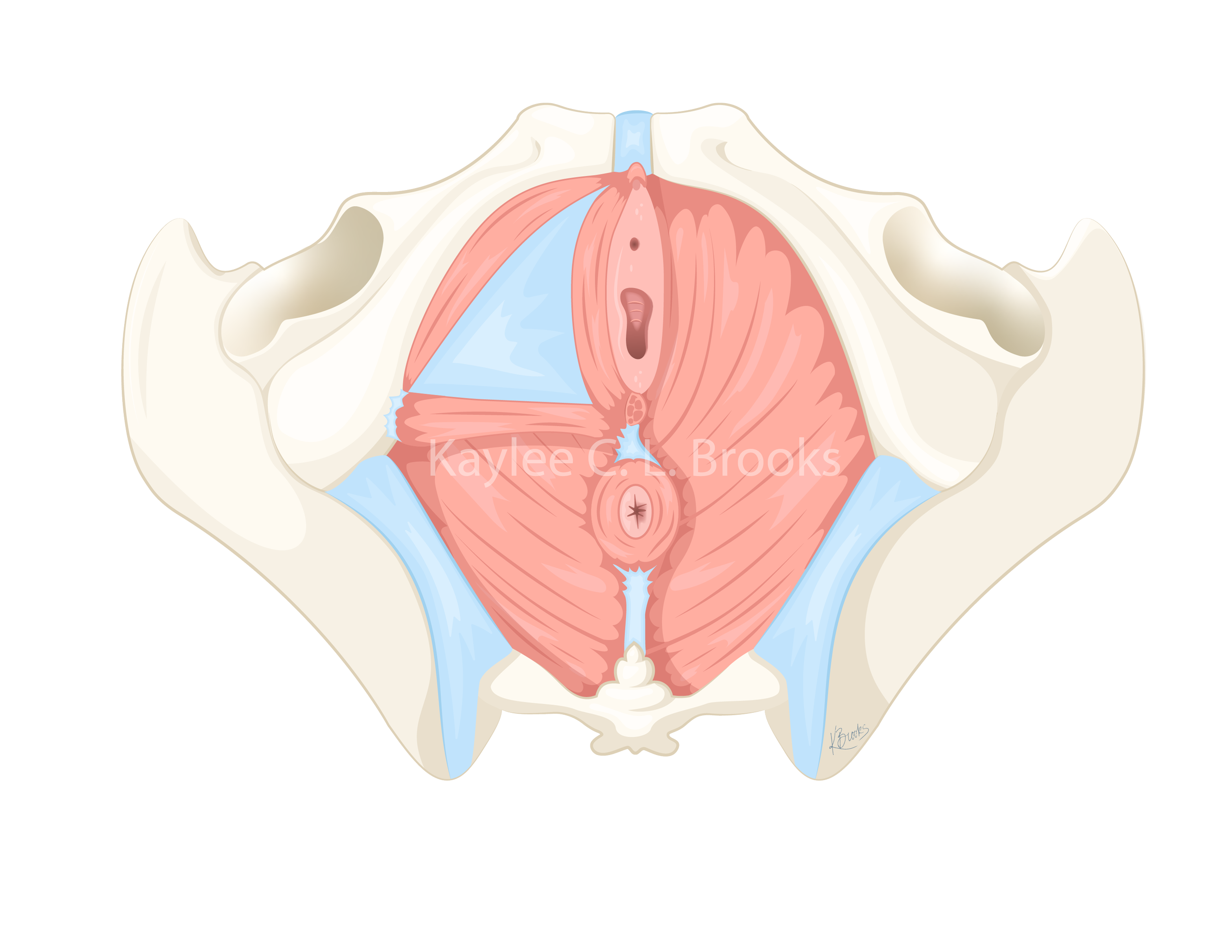
Inferior View of the female pelvic floor muscles. The left superficial muscles have been removed to see the deep pelvic floor muscles. Illustrated using Adobe Illustrator.

Figure demonstrating a hypopressive exercise with proper form in standing. Illustrated using Adobe Illustrator for the MFM Lab Hypopressive Study.
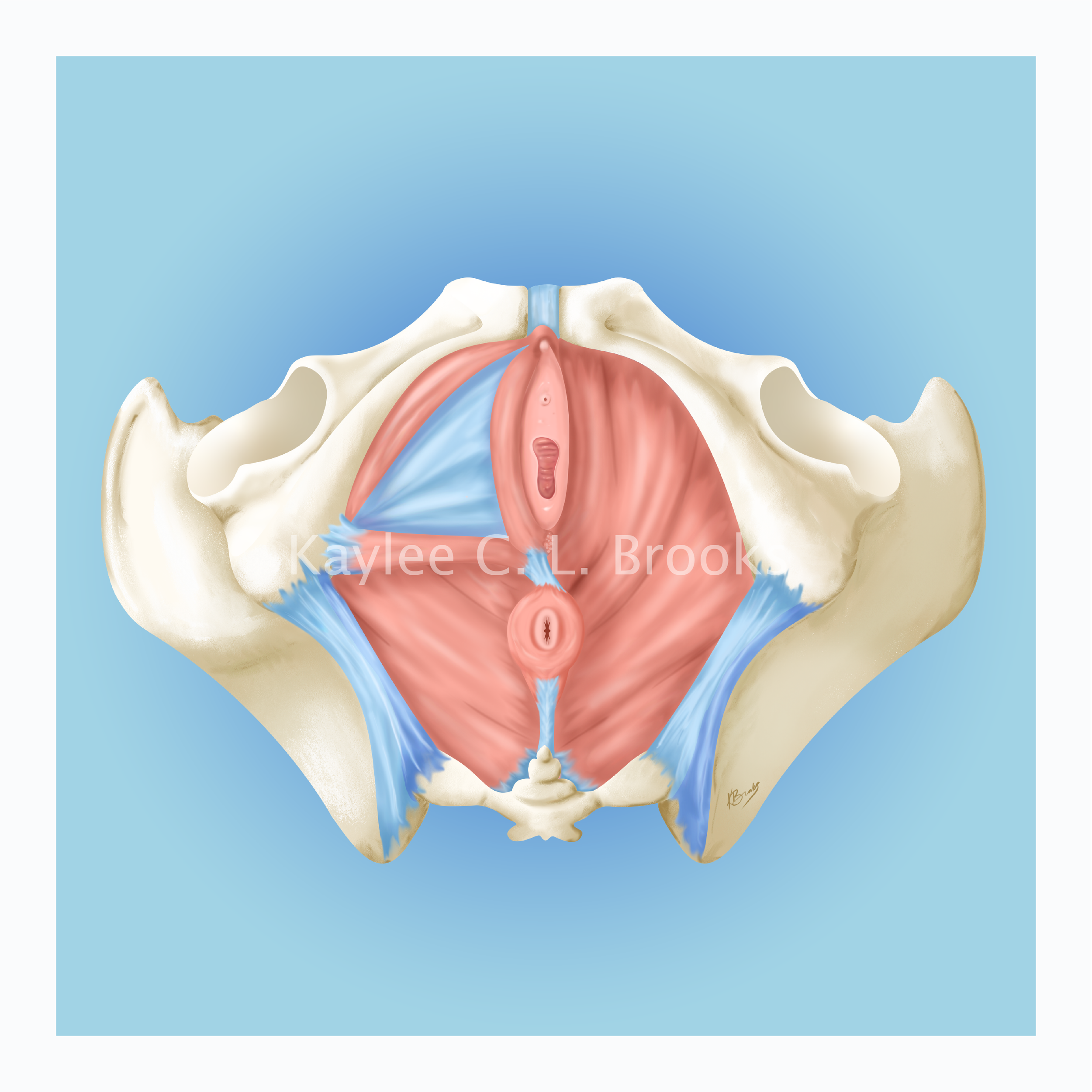
Inferior View of the female pelvic floor muscles. The left superficial muscles have been removed to see the deep pelvic floor muscles. Illustrated using Adobe Illustrator and Fresco (ipad app).
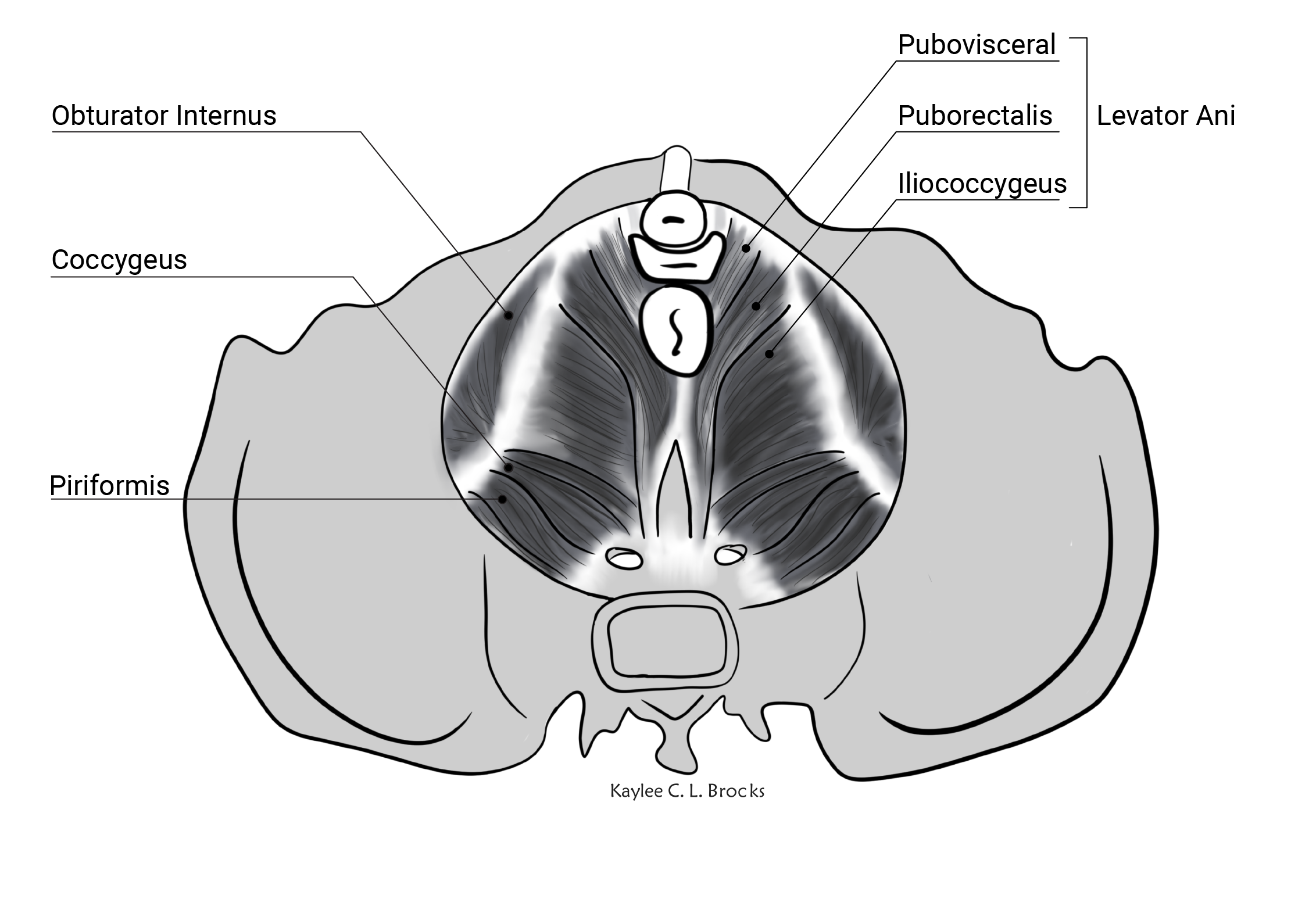
Deep pelvic floor muscles in a top down view illustrated using Krita and Adobe Illustrator.

Intravaginal Dynamometer from the MFM Lab. Illustrated using Adobe Illustrator.
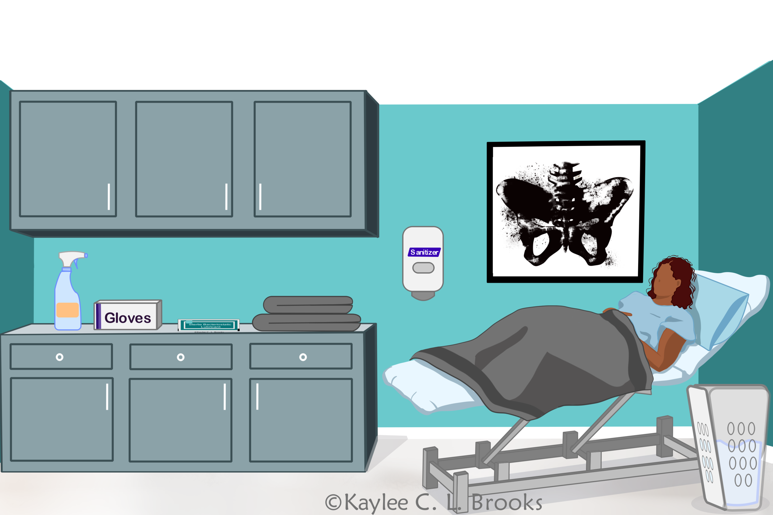
Pelvic floor physiotherapy exam room with a patient displaying appropriate infection control measures in the room. Illustrated using Krita.

Woman during a pelvic floor physiotherapy treatment. Illustrated using Krita.

Curvilinear Ultrasound Probe illustrated using Adobe Illustrator.

Depicting a female experiencing pelvic health issues. Illustrated in Krita.